12. CT Scanners: Summary
ND320 C3 L1 12 CT Scanners- Exercise Summary, Hounsfield Units
Summary
In this module, we have covered the basics of the operation of CT scanner and have gone through an exercise that lets us take a glimpse at what is actually happening when a CT scanner is reconstructing an image.
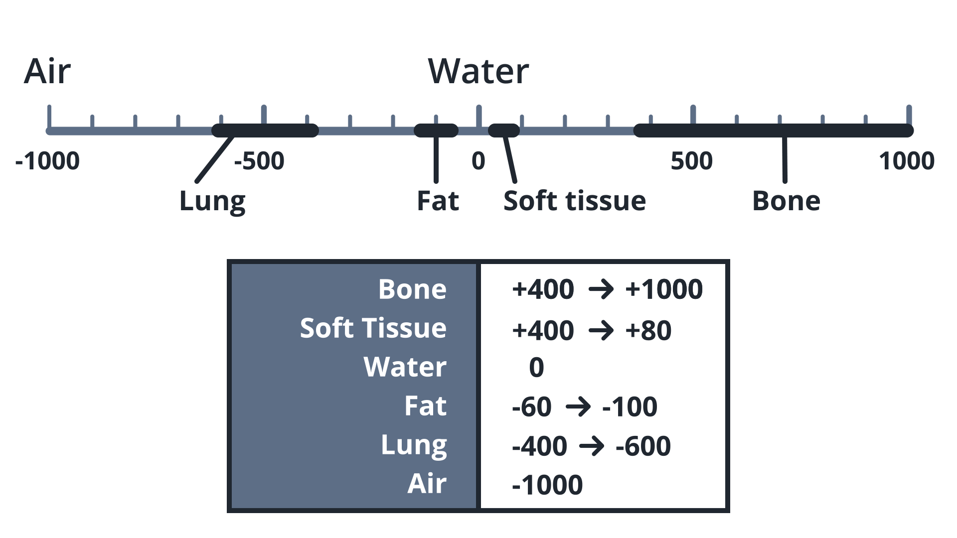
You have seen how the nature of CT data acquisition - the fact that we are effectively measuring radiodensity of biological material - allows us to define a consistent way of associating intensities of pixels in a CT scan slice with the physical density of a structure that is being imaged.
This allows us to guarantee a great degree of consistency among all CT scanners, and thus allow us to have the Hounsfield Scale, named after Sir Godfrey Hounsfield who invented modern CT scanners in the 1970s.
Hounsfield Scale maps tissue types to pixel values of CT scans and is essential to understanding CT scans.
HU scale

Hounsfield Unit Scale
CT Scanners - knowledge check
SOLUTION:
Amount of photons passing through the body attenuated by material between the emitter and detectorSummary 2
If you want to understand the physics of CT scanners a bit further, let me point you to some interesting resources.
Further Resources
Wikipedia offers a very comprehensive description of how CT scanners work, it’s a great resource to get the next level of depth.
An excellent resource that covers some of the basics of CT physics in more depth, as well as offers a little backprojection simulator: http://xrayphysics.com/ctsim.html
A paper on using deep learning methods directly on sinograms: Man, Quinten De, et al. “A Two-Dimensional Feasibility Study of Deep Learning-Based Feature Detection and Characterization Directly from CT Sinograms.” American Association of Physicists in Medicine (AAPM), 2019
A playlist of YouTube videos on CT physics for physicians from the University of Queensland: https://www.youtube.com/watch?v=lbn-IMjF_Uc&list=PL_iaBDDMPsLdxqNDMgAe7ceBMn5FSfpk3